Invertebrate Morphogenesis
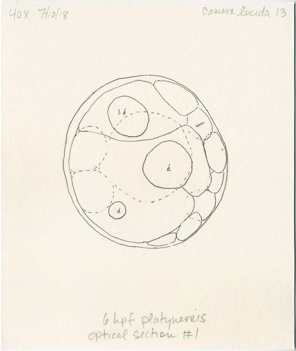
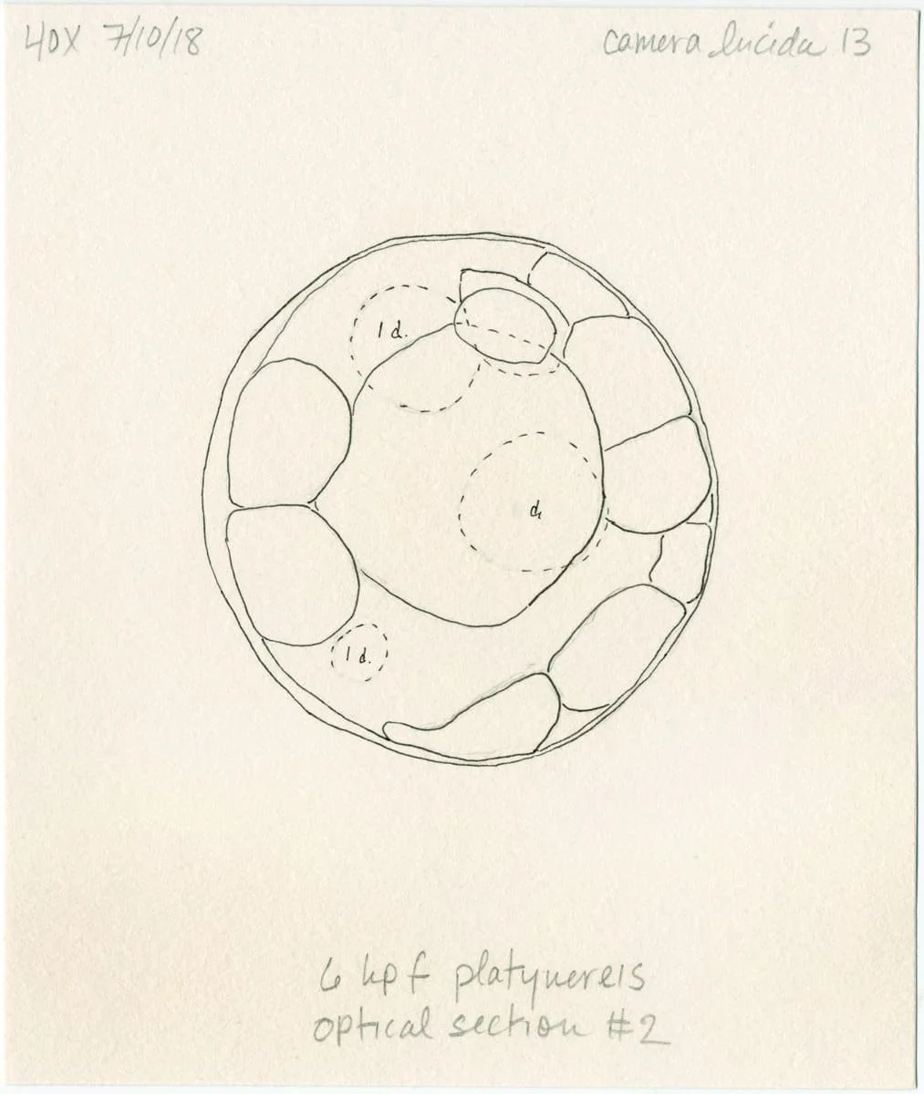
Most of the organisms we encounter on a daily basis emerged from a single cell — an egg, seed, or spore — or small cluster of cells. My observation-intensive research in the lab investigates how cells create increasingly complex shapes in invertebrate animal embryos and larvae. I look at how this process emerges from cell and organelle shape change, movement, and orientation as well as the formation of cell lineage domains and cytoskeletal structures.
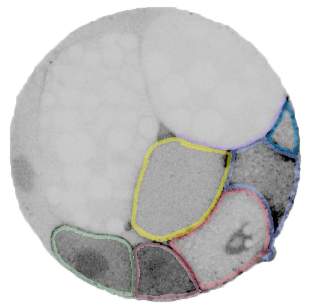
Segmentation of an 32-cell Platynereis dumerilii embryo. These microscope images were used to create the 3D model of the same embryo above.
I am fascinated by how what we can know about the natural world is inseparable from how we see it. At the microscopic scale, we have no other way to know organisms other than through lens-based instrumentation, significant manipulation of the tissue, and some form of image-making. A drawing of a living unstained embryo, a time-lapse sequence of a GFP-labeled tissue, or an animation of a 3D rendering, for example, each provides a different way of knowing life at this scale.
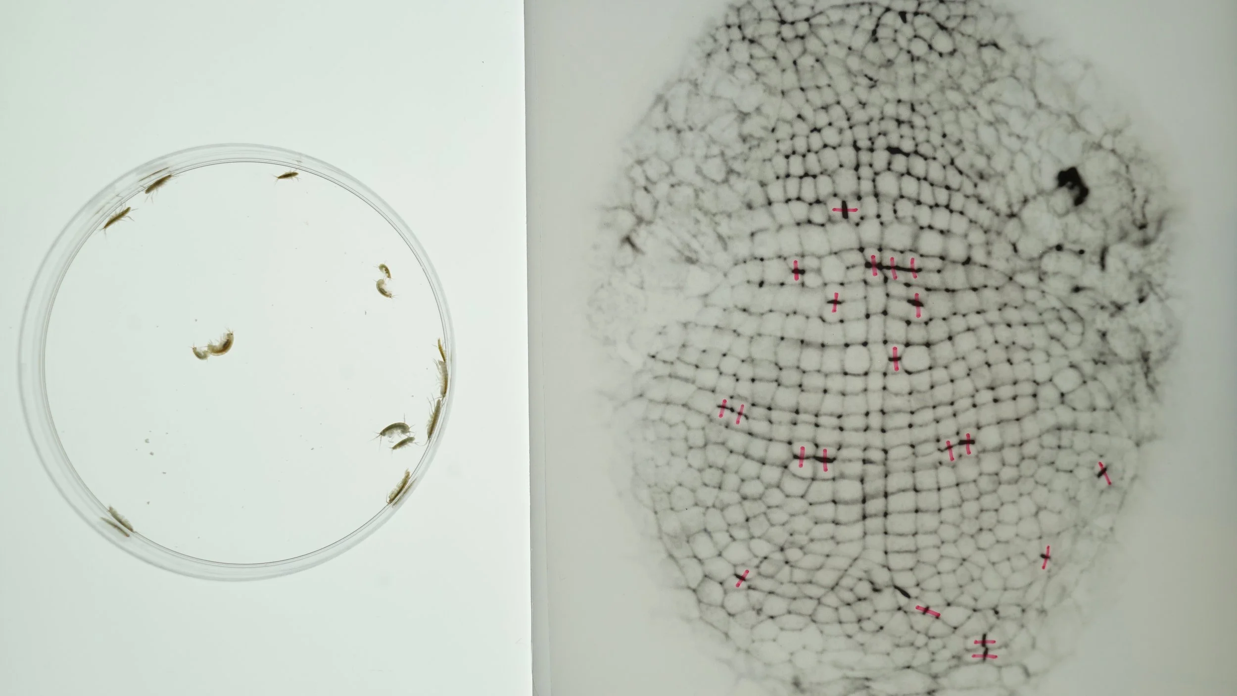
Left: Adult Parhyale hawaiensis. Right: A square grid of cells in the Parhyale hawaiensis embryo (approx. 500 micrometers long). Cells labeled with pink marks are in the process of dividing.
Much of developmental biology involves grappling with the nuances of microscopic change over time and through cellular space. I love thinking about how to capture, comprehend, and represent the movement of cells or sub-cellular structures. My current project focuses on how these transformations contribute to morphogenesis, the development of organismal form, in the embryo of the marine amphipod Parhyale hawaiensis.
Cellular development in the Parhyale germ band, the embryonic structure that establishes the blueprint for the adult body. On the left is a 16-cell stage embryo and on the right is the same embryo approximately two days later.

Movement of neuronal mitochondria in a Drosophila melanogaster larva as a time-lapse animation (left) and kymograph (right). I produced these images to visualize the effect of neurodegenerative mutations on mitochondria morphogenesis in sensory neurons.
I have found that while computational image processing now allows us to see these processes in novel and exciting ways, it is also in many ways upending the embodied observational practices that have been central to biology for centuries. To address this, I am exploring ways of using virtual reality to engage with microscope image datasets.
Visualizing Crepidula fornicata embryos in VR and under the microscope
Video produced by Hyacinth Empinado and STAT News (2017)
In 2018, I co-organized the Embryo Journal Club at the Marine Biological Laboratory in Woods Hole, MA. Our aim was to rediscover classical studies on marine invertebrate development and explore new approaches to addressing old questions. We made our process and findings available through our blog and twitter.